What does fact checked mean?
At Healthfully, we strive to deliver objective content that is accurate and up-to-date. Our team periodically reviews articles in order to ensure content quality. The sources cited below consist of evidence from peer-reviewed journals, prominent medical organizations, academic associations, and government data.
The information contained on this site is for informational purposes only, and should not be used as a substitute for the advice of a professional health care provider. Please check with the appropriate physician regarding health questions and concerns. Although we strive to deliver accurate and up-to-date information, no guarantee to that effect is made.
What Parts of the Brain Produce Dreams?
When Sigmund Freud began investigating dreams 100 years ago, he assumed that dreaming involved many parts of the brain. While the modern science of dreaming has disproved much of Freudian theory, neuroscientists widely accept his central premise that dreams are meaningful expressions of the mind-brain system. The lower, middle and higher brain all contribute to dreaming cognition, making dreams a weird but fruitful object of study.
Lower Brain Causes REM sleep
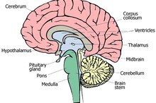
The oldest part of the brain, shared by all vertebrates, is the brain stem. In 1977, Allan Hobson and R McCarley discovered that electrochemical pulses from the brain stem create the stage of sleep in which most dreams occur. Known as REM, which stands for rapid eye movement, this stage of sleep guides the paralysis of all voluntary muscle groups, except for the eyes. Scientists believe these brain pulses from the pons region of the brain stem may create the seemingly random shifts in dream scenery for which dreams are so well known.
- The oldest part of the brain, shared by all vertebrates, is the brain stem.
- In 1977, Allan Hobson and R McCarley discovered that electrochemical pulses from the brain stem create the stage of sleep in which most dreams occur.
Middle Brain Adds Emotions
Parts of the Brain That Influence Creativity
Learn More
When dreaming sleep begins, the middle brain “lights up” with activity. In fact, this part of the brain, which humans share with all mammals, is more activated than in waking life. Also known as the limbic system, the middle brain controls emotional responses and cravings. One organ in the brain is especially active: the amygdala, a walnut-sized mass that philosopher Rene Descartes once thought was the seat of the soul. Today, the amygdala is better called the seat of fear, due to its role in maintaining fight-or-flight responses.
Dream researcher Rosalind Cartwright suggests that dreams are so emotional because we are replaying old memories and updating them with information from recent experiences. It’s not straightforward reason but an emotional kind of logic that links all these memories together. Cartwright’s laboratory research indicates that most dreams are negative in emotion. The most prominent emotional themes in dreams are fear, anxiety, anger and confusion, providing support for the amygdala's role in the dreaming brain.
- When dreaming sleep begins, the middle brain “lights up” with activity.
- The most prominent emotional themes in dreams are fear, anxiety, anger and confusion, providing support for the amygdala's role in the dreaming brain.
Higher Brain Makes Sense of it All
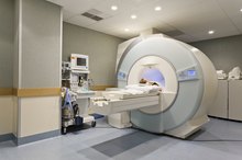
Why don’t we realize when dreaming that monsters, ghosts and goblins are not real? In 2002, co-author Allen Braun of the National Institutes of Health published positron emission tomography, or PET, data from the brain scans of dreaming patients clearly showing how the higher brain is largely offline during dreaming sleep. Specifically, the prefrontal cortex that generates language, logic and critical thinking is taking an electrochemical nap while we run away from our nightmare goblins. However, some critical thinking still occurs in dreams, evidenced by the way we create new outcomes in dreams by trying to “work around” the weird plot changes and bizarre visual imagery.
An exception to the lack of executive functioning in REM sleep may be lucid dreaming, which is when the dreamer knows he is dreaming 3. Validated in the laboratory by Stanford psychophysiologist Stephen LaBerge, lucid dreaming is marked by conscious choices, active thinking and logical reasoning in the dream 3. This claim is strengthened by researcher Ursula Voss, who along with her colleagues from the Neurological Laboratory in Frankfurt, Germany, revealed that the brain has heightened activity in the frontal and frontolateral areas during these “self-aware” dreams.
The science of dreaming is still in its infancy, but neuroscience has come a long way since Dr. Freud in explaining which parts of the brain create dreams.
- Why don’t we realize when dreaming that monsters, ghosts and goblins are not real?
- In 2002, co-author Allen Braun of the National Institutes of Health published positron emission tomography, or PET, data from the brain scans of dreaming patients clearly showing how the higher brain is largely offline during dreaming sleep.
Related Articles

Parts of the Brain That Influence Creativity
Learn More
Parts of the Brain Involved With Hearing
Learn More
What the Different Parts of the Brain Do?
Learn More

Parts of the Brain Used While Driving
Learn More

What Do the Parts of the Brain Control?
Learn More

Parts of the Brain Involved in Memory
Learn More
Four Main Parts of the Brain
Learn More

What Are the Causes of a Sociopath?
Learn More
What Parts of the Brain Control the Parts of Your Body?
Learn More

What Are the Causes of Low Dopamine Levels?
Learn More
References
- Psychology Today: Dreaming up a good mood
- Brain, "The process of awakening," Balkin, Braun, Wesensten, Jeffries, Varga, Baldwin, Belensky, Herscovitch, 2002, 125, 2308-2319
- PubMed: Lucid dreaming
- Ruby PM. Experimental research on dreaming: state of the art and neuropsychoanalytic perspectives. Front Psychol. 2011;2:286. doi:10.3389/fpsyg.2011.00286
- National Institute of Neurological Disorders and Stroke. Brain Basics: Understanding Sleep. Updated August 13, 2019.
- De Gennaro L, Cipolli C, Cherubini A, et al. Amygdala and hippocampus volumetry and diffusivity in relation to dreaming. Hum Brain Mapp. 2011;32(9):1458-70. doi:10.1002/hbm.21120
- Horikawa T, Tamaki M, Miyawaki Y, Kamitani Y. Neural decoding of visual imagery during sleep. Science. 2013;340(6132):639-42. doi:10.1126/science.1234330
- Zhang W, Guo B. Freud's dream interpretation: a different perspective based on the self-organization theory of dreaming. Front Psychol. 2018;9:1553. doi:10.3389/fpsyg.2018.01553
- Wegner DM, Wenzlaff RM, Kozak M. Dream rebound: the return of suppressed thoughts in dreams. Psychol Sci. 2004;15(4):232-6. doi:10.1111/j.0963-7214.2004.00657.x
- Hobson JA, McCarley RW. The brain as a dream state generator: an activation-synthesis hypothesis of the dream process. Am J Psychiatry. 1977;134(12):1335-48. doi:10.1176/ajp.134.12.1335
- Eichenlaub JB, Van Rijn E, Gaskell MG, et al. Incorporation of recent waking-life experiences in dreams correlates with frontal theta activity in REM sleep. Soc Cogn Affect Neurosci. 2018;13(6):637-647. doi:10.1093/scan/nsy041
- Zhang W. A supplement to self-organization theory of dreaming. Front Psychol. 2016;7. doi:10.3389/fpsyg.2016.00332
- Rasch B, Born J. About sleep's role in memory. Physiol Rev. 2013;93(2):681-766. doi:10.1152/physrev.00032.2012
- Marzano C, Ferrara M, Mauro F, et al. Recalling and forgetting dreams: theta and alpha oscillations during sleep predict subsequent dream recall. J Neurosci. 2011;31(18):6674-83. doi:10.1523/JNEUROSCI.0412-11.2011
- Stickgold R, Walker MP. Sleep-dependent memory triage: evolving generalization through selective processing. Nat Neurosci. 2013;16(2):139-45. doi:10.1038/nn.3303
- Llewellyn S, Desseilles M. Editorial: Do both psychopathology and creativity result from a labile wake-sleep-dream cycle?. Front Psychol. 2017;8:1824. doi:10.3389/fpsyg.2017.01824
- Gujar N, McDonald SA, Nishida M, Walker MP. A role for REM sleep in recalibrating the sensitivity of the human brain to specific emotions. Cereb Cortex. 2011;21(1):115-23. doi:10.1093/cercor/bhq064
- Blagrove M, Hale S, Lockheart J, Carr M, Jones A, Valli K. Testing the empathy theory of dreaming: the relationships between dream sharing and trait and state empathy. Front Psychol. 2019;10:1351. doi:10.3389/fpsyg.2019.01351
- Kets de Vries MFR. Dream journeys: a new territory for executive coaching. Consulting Psychology Journal: Practice and Research. 2014;66(2):77–92. doi:10.1037/cpb0000004
- Baird B, Castelnovo A, Gosseries O, Tononi G. Frequent lucid dreaming associated with increased functional connectivity between frontopolar cortex and temporoparietal association areas. Sci Rep. 2018;8(1):17798. doi:10.1038/s41598-018-36190-w
- Vallat R, Ruby PM. Is it a good idea to cultivate lucid dreaming?. Front Psychol. 2019;10:2585. doi:10.3389/fpsyg.2019.02585
- Baird B, Mota-Rolim SA, Dresler M. The cognitive neuroscience of lucid dreaming. Neurosci Biobehav Rev. 2019;100:305-323. doi:10.1016/j.neubiorev.2019.03.008
- Stumbrys T, Daunytė V. Visiting the land of dream muses: The relationship between lucid dreaming and creativity. 2018;11(2). doi:10.11588/ijodr.2018.2.48667
- Rek S, Sheaves B, Freeman D. Nightmares in the general population: Identifying potential causal factors. Soc Psychiatry Psychiatr Epidemiol. 2017;52(9):1123-1133. doi:10.1007/s00127-017-1408-7
- Sikka P, Pesonen H, Revonsuo A. Peace of mind and anxiety in the waking state are related to the affective content of dreams. Sci Rep. 2018;8(1):12762. doi:10.1038/s41598-018-30721-1
Resources
Writer Bio
Ryan Hurd is a writer and consciousness studies researcher living in California. His dream expertise has been featured in the Huffington Post and Psychology Today. Hurd has an M.A. in consciousness studies, and is the author of "Enhance your dream life."