Tendonitis Symptoms in Hands
Finger movements are controlled by tendons -- connective tissue cords that attach your finger bones to muscles in your forearms. Overloading your finger tendons by lifting an item that is too heavy, or lifting too quickly can cause inflammation in these tendons, a condition broadly called tendinitis. Microtears may also develop in the tendon. Overuse of the muscles that move your fingers may also cause inflammation and pain. This type of injury is also often called tendinitis, however according to a study published in 2012 by the "International Journal of Therapeutic Massage & Bodywork Consult", it is better described as tendinopathy. See your doctor for an accurate diagnosis if you experience symptoms of finger tendinitis.
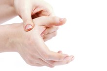
Pain and Swelling
Hand tendinitis is a painful condition. Pain may be sharp, achy or you may feel a burning sensation. Pain increases as you use your hand, and typically decreases with rest. Tendinitis commonly develops in the tendons that bend your fingers. You may feel pain on the front of your finger, or along the length of the tendon as it travels through your palm, particularly with activities such as gripping and typing. Direct pressure on the tendon -- such as holding a pen -- can also be painful. Swelling may develop over the tendon, causing a small puffy area on your finger or palm.
- Hand tendinitis is a painful condition.
- Swelling may develop over the tendon, causing a small puffy area on your finger or palm.
Popping or Catching
Hand Tendon Exercises
Learn More
Finger tendons glide through tunnels in your hand as your fingers move. Tendinitis may cause finger tendons to swell, making it difficult for the tendons to glide through these tunnels. Severe tendinitis can cause a condition called trigger finger -- the tendon of the affected finger becomes caught or stuck in the tunnel as the finger bends. As you straighten your finger, a painful popping sensation occurs as the tendon glides through the tunnel. In severe cases, you may have to straighten the affected finger using your other hand. According to a study published in 2012 by "Clinics in Orthopedic Surgery", trigger finger is one of the most common diagnoses seen by hand surgeons. See your doctor if you experience these symptoms -- medical treatment may be required.
- Finger tendons glide through tunnels in your hand as your fingers move.
- As you straighten your finger, a painful popping sensation occurs as the tendon glides through the tunnel.
Weakness and Stiffness
Inflammation caused by hand tendinitis may affect your hand strength and ability to move your fingers. Hand activities such as gripping a doorknob, removing a container of milk from the fridge or holding a pen are often difficult. You may even drop objects. Prolonged symptoms can also lead to stiffness in your finger joints, affecting your ability to make a tight fist or fully straighten your fingers. Early treatment for finger tendinitis reduces your risk of developing these symptoms.
- Inflammation caused by hand tendinitis may affect your hand strength and ability to move your fingers.
- Prolonged symptoms can also lead to stiffness in your finger joints, affecting your ability to make a tight fist or fully straighten your fingers.
Related Articles
Hand Tendon Exercises
Learn More

Rehabilitation Exercises for a Dislocated Finger
Learn More

Military Press and Elbow Tendons
Learn More

Carpal Tunnel Syndrome After Childbirth
Learn More

Dupuytren's Contracture Exercises
Learn More

Trigger Finger Exercises
Learn More
Symptoms of a Torn Tendon in the Big Toe
Learn More

Old-Fashioned Remedies for Stiff Finger Tendons
Learn More
Signs of a Torn Ligament in the Finger
Learn More

Ankle Tendinitis Symptoms and Treatment
Learn More
References
- Clinics in Orthopedic Surgery: The Efficacy of Steroid Injection in the Treatment of Trigger Finger
- Journal of Ultrasound: Stenosing Tenosynovitis
- International Journal of Therapeutic Massage & Bodywork: Tendinopathy: Why the Difference Between Tendinitis and Tendinosis Matters
- American Academy of Orthopaedic Surgeons. Mallet Finger (Baseball Finger). Updated March 2015.
- Pingel C, McDowell C. Subungual hematoma drainage. StatPearls Updated May 18, 2019.
Writer Bio
Rae Uddin has worked as a freelance writer and editor since 2004. She specializes in scientific journalism and medical and technical writing. Her work has appeared in various online publications. Uddin earned her Master of Science in integrated biomedical sciences with an emphasis in molecular and cellular biochemistry from the University of Kentucky College of Medicine.