How Big Is My Unborn Baby at 18 Weeks?
Pregnancy is said to last about 40 weeks and is divided into three trimesters. Week 18 falls in the second trimester and is near the halfway point, 20 weeks.
The second trimester is sometimes called the "honeymoon" period since it is a more comfortable trimester of pregnancy 1. This is because the woman has likely overcome morning sickness and fatigue. At 18 weeks along, an unborn baby begins resembling a human being and is developing some identifiable characteristics, including genitalia.
Mother's Bodily Changes
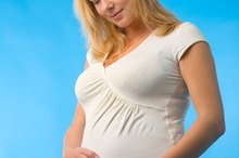
At 18 weeks pregnant a woman has gained some weight and her body has changed shape in various ways. MayoClinic.com suggests a woman's breasts may be larger and her belly may be visibly larger. First-time mothers may be starting to show slightly and women who have had previous pregnancies will likely have a noticeably expanded waistline, often called a baby bump. The actual weight a woman puts on by the 18th week is difficult to predict. Some women may not gain much by this point while others gain an average of half a pound per week. The care provider working with the mother can help set weight gain goals to ensure the mother gains enough to support herself and the growing infant. By this point in the pregnancy the weight a mother has gained is only slightly due to the weight of the unborn baby.
- At 18 weeks pregnant a woman has gained some weight and her body has changed shape in various ways.
- The actual weight a woman puts on by the 18th week is difficult to predict.
Unborn baby's Weight
Normal Endometrial Stripe Thickness
Learn More
MayoClinic.com states that by the 20th week a baby may weigh about 9 ounces 1.
Predicting the exact weight of a baby at 18 weeks is difficult because of various factors including genetics and the mother's diet.
The American Congress of Obstetricians and Gynecologists suggests that at 16 weeks along a baby is likely to be six or seven inches long and weigh about 5 ounces. Some women struggle to eat well during the first trimester, when morning sickness is most prominent.
- MayoClinic.com states that by the 20th week a baby may weigh about 9 ounces 1.
- The American Congress of Obstetricians and Gynecologists suggests that at 16 weeks along a baby is likely to be six or seven inches long and weigh about 5 ounces.
Baby's Development
In addition to putting on a few ounces, the baby has developed many new skills including the ability to kick, make facial expressions and hear states MayoClinic.com. The American Congress of Obstetricians and Gynecologists indicates that by the fourth month the baby has developed eyebrows, eyelashes, fingernails, external sex organs, skin wrinkles, outer ears, a neck and the kidneys are beginning to produce urine.
Ultrasound
Signs of Re-Canalization From a Vasectomy
Learn More
A baby's growth is measured at routine prenatal appointments, typically conducted every four weeks until the third trimester. Some providers conduct an ultrasound around the 18th or 20th week of pregnancy.
An ultrasound allows a qualified technician to take measurements of the baby's head, crown-to-rump height and various other points to estimate the baby's weight and age. The crown-to-rump height is the length from the top of the baby's head to the base of the tailbone or visible bottom.
This measurement is taken because the baby is usually curled up tight, called the fetal position. DrSpock.com explains that these measurements are compared to a chart to estimate the baby's development and age.
Another way caregivers can estimate a baby's growth and the progress of the pregnancy is to measure the fundal height. MayoClinic.com explains that this is the distance from the top of the uterus to the pubic bone. The woman's fundal height is compared to a chart to determine whether or not the pregnancy is progressing normally.
- A baby's growth is measured at routine prenatal appointments, typically conducted every four weeks until the third trimester.
- DrSpock.com explains that these measurements are compared to a chart to estimate the baby's development and age.
Considerations
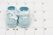
Various factors can contribute to the accuracy of measuring a baby's weight at 18 weeks into the pregnancy. In the beginning a woman's estimated due date is often based on her last menstrual period. From this date a caregivers estimates the due date to be 40 weeks. There are various ways this estimate can be inaccurate, including the woman's period starting sooner or later than she recalls or notices.
Ultrasounds performed on or before the 20th week can help in properly dating the pregnancy but there is room for error in these measurements as well. MayoClinic.com points out that measuring the fundal height can lead to an inaccurate estimate if the woman has a tall or slim frame, well-conditioned abdominal muscles or the baby has descended into the pelvic region early in the pregnancy 12. These factors aren't harmful but they can contribute to a smaller or larger estimate of the baby's weight. Continued prenatal care allows caregivers to monitor such measurements. If there is a problem, the measurements will fluctuate suggesting the baby is due sooner or later than the previous measurements. If the baby is continually measuring ahead or behind, the due date may be adjusted under the assumption that the original estimated due date was incorrect.
- Various factors can contribute to the accuracy of measuring a baby's weight at 18 weeks into the pregnancy.
- Ultrasounds performed on or before the 20th week can help in properly dating the pregnancy but there is room for error in these measurements as well.
Related Articles
Normal Endometrial Stripe Thickness
Learn More

Signs of Re-Canalization From a Vasectomy
Learn More
Normal Levels of HCG & Progesterone
Learn More

The Average Weight & Height for a 13-Year-Old
Learn More

The Process of Human Reproduction
Learn More

The Average Body Weight for Children
Learn More
How to Work Out Conception Date From Birth Date
Learn More
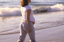
Can Walking Help a Baby Drop?
Learn More

The Size of a 24-Week-Old Baby
Learn More

How to Use a Balance Beam Scale
Learn More
References
Writer Bio
Sarah Harding has written stacks of research articles dating back to 2000. She has consulted in various settings and taught courses focused on psychology. Her work has been published by ParentDish, Atkins and other clients. Harding holds a Master of Science in psychology from Capella University and is completing several certificates through the Childbirth and Postpartum Professional Association.