Can I Eat Before an MRI Brain Scan?
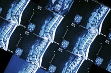
Magnetic resonance imaging, or MRI, is a noninvasive device used for evaluation, assessment and diagnosis of conditions, diseases and injuries. An MRI generates images of the brain and spine from multiple angles, and it also can provide pictures of blood vessels, tissues and organs. The images captured can reflect abnormalities in structure and function within the brain and entire body. The diagnostic value of the MRI scan, invented in 1977, is both precise and detailed in its accuracy.
Function
The MRI scanner uses magnetic field and radio-frequency energy to create pictures based on the hydrogen atom capacity in body tissues. Hydrogen atoms, which are essentially water, are the most plentiful and magnetically susceptible atoms in the body. The magnet pulls the hydrogen atoms into a line, radio waves are directed at the line and sends images to the MRI computer. These images reflect the consistency of the bones, tissues and vessels scanned.
- The MRI scanner uses magnetic field and radio-frequency energy to create pictures based on the hydrogen atom capacity in body tissues.
Preparation
Side Effects of Novocain
Learn More
Before the test, a requirement to fast from food and beverages is based on particular exams and facility policies. Fear of enclosed spaces or excessive anxiety could require sedation for relaxation and to minimize movement. The use of sedation can cause vomiting with possible aspiration of stomach contents, making it necessary to fast. If your scan requires oral or intravenous contrast material, fasting requirements could apply before the exam. Follow your physician's MRI scan instructions.
- Before the test, a requirement to fast from food and beverages is based on particular exams and facility policies.
- The use of sedation can cause vomiting with possible aspiration of stomach contents, making it necessary to fast.
Restrictions
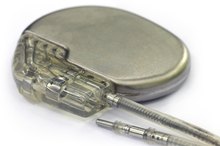
There are instances when an MRI is not performed because of the relationship between the magnetic field and implanted devices or metallic injuries. A few examples include brain aneurysm repair clips, synthetic heart valves, inner ear implants, pacemakers, gunshot wounds, shrapnel and metallic joint replacements. Sheet metal workers and welders might require skull x-rays to detect metal pieces before an MRI. Ventilators and computerized or metal equipment are prohibited from the magnetic field area.
- There are instances when an MRI is not performed because of the relationship between the magnetic field and implanted devices or metallic injuries.
Interference
Side Effects of a Bone Scan
Learn More
Excessive movement can blur the images from the MRI scan. Dental fillings, implants and braces can hinder and distort skull pictures. Some tattoo dyes contain iron and can become very hot and cause serious burns. Medication patches, which are designed to release a drug over an extended period of time, frequently contain aluminum that also can heat and burn the individual. Remember to inform lab personnel of any of these pre-existing circumstances.
- Excessive movement can blur the images from the MRI scan.
- Dental fillings, implants and braces can hinder and distort skull pictures.
Post-test Evaluation
Depending on your particular exam, the scan can take 45 minutes to an hour or more. A radiologist, who is a medical doctor specifically trained to oversee and evaluate your test, notifies your physician of the results. You can restart medications, resume activities and return to your previous diet if fasting was required. An uncommon complication called nephrogenic systemic fibrosis occurs with large doses of contrast material administered to individuals with kidney disorders.
- Depending on your particular exam, the scan can take 45 minutes to an hour or more.
Related Articles
Side Effects of Novocain
Learn More
Side Effects of a Bone Scan
Learn More

Foods Allowed Before a Thyroid Test
Learn More

Why High Levels of Potassium in Burn Patients?
Learn More

Lexapro & Elevated Liver Enzymes
Learn More
Allergic Reaction to Barium Sulfate
Learn More

Four Components of Nutrition Assessment
Learn More

Pros & Cons of X-Rays
Learn More
Side Effects of Exercise With a Cardiac Pacemaker
Learn More
Arthrogram Side Effects
Learn More
References
Resources
Writer Bio
Helen Messina started writing in 2010. She is a registered nurse with experience in rehabilitation, long-term/subacute care, pediatric/adult home care and has worked in acute care facilities in Florida, Pennsylvania, New Jersey and New York. Messina's specialties include neurology, cardiac and renal care. She holds an associate degree in nursing from Gannon University.