Bone Cancer of the Tibia Symptoms
Bone cancer can affect any bone in the body. The tibia is the larger of the two long bones between the knee and ankle. A cancerous tumor is a body of tissue consisting of cells that have undergone genetic changes, causing them to grow in an uncontrolled way. A tibial tumor can be a primary bone tumor -- meaning it originated in the bone -- or a metastatic bone tumor that has spread from cancer elsewhere in the body. Common cancer sites that metastasize to bone include the prostate, thyroid, breast, lung and kidney.
If you are experiencing serious medical symptoms, seek emergency treatment immediately.
Pain
Pain is the most common symptom of bone cancer, although not all people experience this symptom. The pain is usually described as dull or achy. It may come and go initially, but become more persistent over time. The pain might occur or be more noticeable at night, and activity may worsen the discomfort. The pain can occur at the site of the tumor or in nearby joints. With a cancerous tumor of the tibia, pain typically occurs in the shin, knee or ankle.
- Pain is the most common symptom of bone cancer, although not all people experience this symptom.
- The pain can occur at the site of the tumor or in nearby joints.
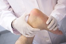
Visible Lump and Swelling
Early Bone Cancer Symptoms
Learn More
The presence of a lump along the shin may be a sign of a tibial tumor. The mass can be painful or painless, depending on the type of tumor and whether it is impinging on nearby structures. For example, a growing tibial tumor can cause nerve compression or bursitis -- painful inflammation of a fluid-filled sac found near body joints to provide cushioning and reduce friction. The growing mass can be firm or soft, and immobile or mobile, depending on the type of tumor. Swelling of the lower leg can also occur with a cancerous tibial tumor.
- The presence of a lump along the shin may be a sign of a tibial tumor.
- The mass can be painful or painless, depending on the type of tumor and whether it is impinging on nearby structures.
Tibia Fracture
Fractures can occur in a bone weakened by a tumor. These fractures, known as pathological fractures, typically occur with minimal or no trauma. As with traumatic bone fractures, a pathological tibia fracture is typically accompanied by sudden intense pain, deformity of the fracture site, and an inability to weight-bear and move the lower leg normally. Pathological tibia fractures can occur near the knee, mid-shin or at the ankle.
- Fractures can occur in a bone weakened by a tumor.
- Pathological tibia fractures can occur near the knee, mid-shin or at the ankle.
General Cancer Symptoms
Hip Bone Cancer Symptoms
Learn More
As with any type of cancer, bone cancer of the tibia can cause generalized symptoms 2. Symptoms may include:
- fevers
- chills
- night sweats
- unexplained fatigue
- changes in appetite
- unintentional weight loss
These symptoms might indicate that the tibial tumor is metastatic and the primary tumor may be located elsewhere in the body 2.
Seek medical care as soon as possible if you experience any signs or symptoms that might indicate a tumor of the tibia. While the possibility of cancer is scary, keep in mind that most bone tumors are not cancerous. Even if cancer is diagnosed, early detection and treatment give you the best chance for a cure.
Reviewed by: Tina M. St. John, M.D.
- Seek medical care as soon as possible if you experience any signs or symptoms that might indicate a tumor of the tibia.
- While the possibility of cancer is scary, keep in mind that most bone tumors are not cancerous.
Related Articles
Early Bone Cancer Symptoms
Learn More
Hip Bone Cancer Symptoms
Learn More

Signs and Symptoms of Bone Cancer in the Thigh
Learn More
What Are the Causes of Pain in the Upper Hip or Waist Area?
Learn More

Signs & Symptoms of Shoulder Injuries
Learn More

Foods That Fight Bone Cancer
Learn More
Cancer & Leg Pain
Learn More
Top Bone Diseases
Learn More

What Are the Signs & Symptoms of Bladder & Kidney Cancer?
Learn More
Signs of a Bone Spur in Elbow
Learn More
References
- AAOS Comprehensive Orthopaedic Review; Jay R. Lieberman, M.D.
- Bone and Soft Tissue Tumors: A Multidisciplinary Review with Case Presentations; Therese J. Bocklage, M.D., et al.
- Merck Manual Professional Version: Primary Malignant Bone Tumors
- Johns Hopkins Medicine Health Library. Tibia and fibula fractures. Date unknown.
- American Academy of Orthopaedic Surgeons. Tibia (shinbone) shaft fractures. Date unknown.
- Patel NK, Horstman J, Kuester V, Sambandam S, Mounasamy V. Pediatric tibial shaft fractures. Indian J Orthop. 2018;52(5):522-528. doi:10.4103/ortho.IJOrtho_486_17
- American Academy of Orthopaedic Surgeons. Fractures of the proximal tibia (shinbone). Date unknown.
- Hofmann A, Dietz SO, Pairon P, Rommens PM. The role of intramedullary nailing in treatment of open fractures. Eur J Trauma Emerg Surg. 2015;41(1):39-47. doi:10.1007/s00068-014-0485-5
- Cereijo C, Attum B, Rodriguez-buitrago A, Jahangir AA, Obremskey W. Intramedullary nail fixation of tibial shaft fractures: Suprapatellar approach. JBJS Essent Surg Tech. 2018;8(3):e24. doi:10.2106/JBJS.ST.17.00063
- American Academy of Orthopaedic Surgeons. Open fractures. Date unknown.
- Raju K, Smith TO, Hing CB, Solan MC, Nielsen DM. Surgical versus conservative interventions for treating tibial shaft fractures in adults. Cochrane Database Syst Rev. 2018;2018(4):CD011095. 2018 April. doi:10.1002/14651858.CD011095.pub2
- Bedi A, Le TT, Karunakar MA. Surgical treatment of nonarticular distal tibia fractures. J Am Acad Orthop Surg. 2006 Jul;14(7):406-16.
- Bono CM, et al. Nonarticular Proximal Tibia Fractures: Treatment Options and Decision Making. J Am Acad Orthop Surg. May/June 2001; 9:176-186.
- Tejwani N, Polonet D, Wolinsky PR. Controversies in the intramedullary nailing of proximal and distal tibia fractures. J Am Acad Orthop Surg. 2014 Oct;22(10):665-73.
Writer Bio
Gregory Waryasz, MD, has been a writer since 2004. His work has appeared in "Dynamic Medicine," "Athletic Therapy Today," the "Strength and Conditioning Journal" and the "Journal of the American Academy of Nurse Practitioners." He is an orthopaedic surgery resident at Brown University/Rhode Island Hospital. He has a medical doctorate from Tufts University and is certified as a strength and conditioning specialist.