


Our internal organ health page highlights the importance of taking care of your body's internal organs. We offer a variety of resources to help you better understand the function of your internal organs, as well as common diseases and conditions that can affect them.
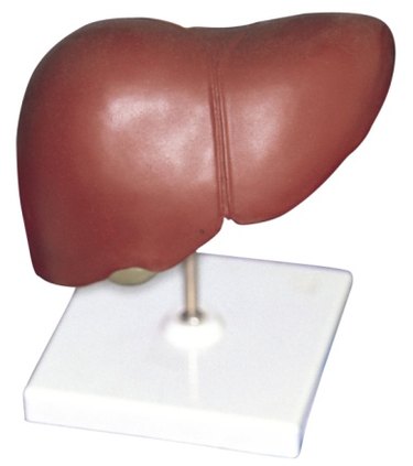
Here you'll find in-depth articles featuring medical experts about specific internal organs, including the heart, lungs, liver, kidneys and digestive system. We also offer advice on how to maintain optimal organ health through lifestyle changes such as diet, exercise and stress reduction.
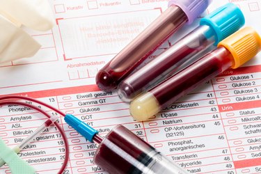
Whether you are dealing with a specific internal organ condition or are looking to improve your overall organ health, our internal organ health page has something for you. We offer practical tips for managing organ conditions, advice on how to prevent organ issues from developing, and information on various organ health resources and services."