


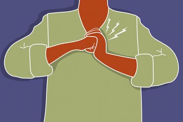


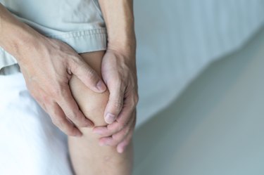


At our joint, ligament and tendon conditions page, we know the effect these issues can have on daily life and mobility. That's why we offer a variety of resources to help you better understand these complex conditions and manage symptoms effectively.
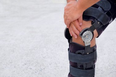
Our team of medical experts provides in-depth articles on specific joint, ligament, and tendon disorders, as well as general information on joint health and function. We also offer practical advice on managing symptoms, including pain, inflammation and limited mobility, and provide information on various treatment options, from medication to therapy and surgery.
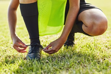
This page is also a great resource for athletes and individuals who engage in physical activity. We offer tips on injury prevention, training strategies and rehabilitation techniques to help you stay active and healthy."