Along with the thermometer as a must-have tool in the time of COVID, pulse oximeters are being snatched up in the hopes of catching serious complications of the illness early. But how do you really use and read a pulse oximeter?
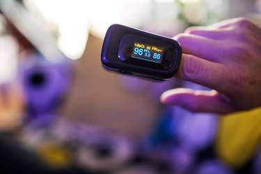
"A pulse oximeter is a small device that uses infrared light to measure how much oxygen is bound to the hemoglobin in your bloodstream," Timothy Connolly, MD, a pulmonologist at Houston Methodist, tells LIVESTRONG.com. "Traditionally, we put them on a patient's finger. It's a wonderful tool to get a sense of their oxygen status."
Video of the Day
Video of the Day
That's where the COVID connection comes in. Among the many symptoms of COVID-19 is shortness of breath. But an at-home pulse oximeter shouldn't be used to diagnose the disease, because by the time you have difficulty breathing, you might already be in trouble.
Get tips on how to stay healthy, safe and sane during the novel coronavirus pandemic.
"What we have seen over the last 10 months is that patients present to the hospital when they feel short of breath. By the time they arrive, they already have evidence of hypoxia — low oxygen levels — and require aggressive interventions, either with high-flow oxygen, BPAP or even intubation," Dr. Coleman says.
In other words, hypoxia often comes before shortness of breath and might not come with any symptoms, either. You might feel "fine," and then decline rapidly. This is called silent hypoxia, and these low oxygen levels in the body can cause life-threatening damage to organs, research indicates. You might also hear this referred to as "happy hypoxia."
This is where a pulse oximeter might come in handy: If you have already been diagnosed with COVID-19 and find that your oxygen levels are declining — even if you don't feel terribly sick — you have an indication that you're not doing well, your condition may decline and you have the green light to go to the hospital.
Tell the personnel that you took your pulse oximeter reading and what it was. Doctors will confirm it and use that info to inform your treatment.
Where Can You Buy a Pulse Oximeter?
These devices are easy to find and inexpensive to buy. They cost as little as $30 (and often can be purchased with FSA dollars) and are found through retailers like Walgreens or on Amazon.com.
How to Use a Pulse Oximeter
- Follow the manufacturer's instructions.
- Put the oximeter on your index, middle or ring finger and make sure it fits well (not too loose, not too tight).
- To get the most accurate reading, allow 5 to 20 seconds for your pulse rate to stabilize and for the oximeter to detect your pulse.
Tip
Most at-home oximeters can determine normal oxygen saturation, but they are not as accurate when levels are below normal. If you have questions about your reading, always discuss with your health care provider.
Everyone has a reading of what's "normal" for them, so you'll want to get a good baseline, Dr. Connolly says.
Ideally, you'll take your reading when you're well, about once per day, Dr. Coleman says. You're looking for a reading in the mid- to high-90s, he says.
If you have COVID, check your oxygen levels twice a day, in the morning and evening. Seek medical attention for levels in the upper 80s, even if you feel as if you can breathe just fine, Dr. Coleman says.
You might also want to check it during times of different activity, such as when you get up to walk to the bathroom or when you're sitting on your couch.
Check on different fingers, too, as factors you wouldn't expect (dark nail polish, fake nails, structure of finger nails) can throw things off, Dr. Connolly says. If you check one finger and it gives you an alarmingly low reading, check a finger on the other hand. If that finger's reading is normal, then you're OK.
If you're worried or have questions about your readings, call your doctor.
What if You Do Not Have a Pulse Oximeter?
If you've been diagnosed with COVID-19 and do not have a pulse oximeter, you can monitor yourself for shortness of breath.
"The typical symptoms of low oxygen or hypoxia are shortness of breath, either at rest or with exertion," Dr. Coleman says, adding that you might notice this walking up or down stairs or generally feeling sleepier during the day.
Read more stories to help you navigate the novel coronavirus pandemic:
Is this an emergency? If you are experiencing serious medical symptoms, please see the National Library of Medicine’s list of signs you need emergency medical attention or call 911.